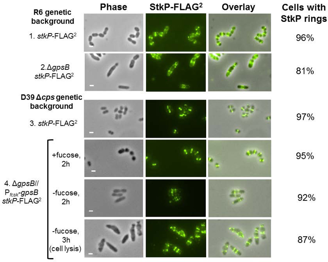
Fig.7.
2D IFM microscopy demonstrates that the absence or depletion of GpsB does not abolish StkP ring formation. 2D IFM was performed as outlined in Experimental procedures. Panels shown from left to right are: phase, FITC antibody labeled FLAG-tagged StkP, and phase/FITC overlay. 1) R6 stkP-FLAG2 (IU8819, sampled at OD620 ≈ 0.2); 2) R6 ΔgpsB stkP-FLAG2 (IU8311, sampled at OD620 ≈ 0.2); 3) D39 Δcps stkP-FLAG2 (IU7434, sampled at OD620 ≈ 0.2); 4) merodiploid strain D39 Δcps stkP-FLAG2 Δcps ΔgpsB//PfcsK-gpsB+ (IU8230) grown for 2 h with fucose addition or without fucose for 2 h or 3 h to deplete GpsB, eventually causing cell lysis. Representative images of each strain are shown for each experiment, which were performed three times independently with similar results. Percentages of cells with StkP rings are based on 100 manually examined cells of each strain. Scale bar = 1 micron.

