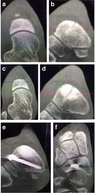
Fig. 1.

Coronal (a) and sagittal (b) weight-bearing CT images demonstrate a type I navicular stress fracture in a division I track athlete. After 6 weeks of non-weight bearing and boot immobilization, the coronal (c) and sagittal (d) weight-bearing CT images demonstrate increased sclerosis and persistence of the fracture line. The patient underwent percutaneous screw fixation with BMAC and CT images (e, f) demonstrate fracture healing at 10 weeks post-operatively
