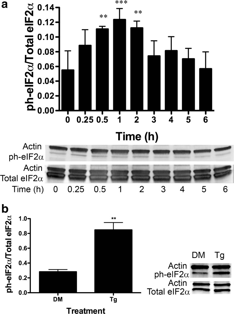
Fig. 2.
BA induces eIF2α phosphorylation at ser-51 in DU-145 cells. a DU-145 cells were treated with 10 μM BA for 0, 0.25, 0.5, 1, 2, 3, 4, 5, and 6 h. Phosphorylation of eIF2α was significantly higher at 0.5 (p < 0.006, n = 3), 1, (p < 0.001, n = 3), and 2 h (p < 0.005, n = 3) post-treatment. b DU-145 cells were treated with 1 μM thapsigargin (Tg), a strong positive control that induces stress and apoptosis, or DMSO (DM), the vehicle for Tg for 1 h, p < 0.01, n = 3. In Figs. 2, 3, 4, 5, 6, 7, 8, 9, 10, 11, and 12, the probabilities of statistical differences are represented as *p < 0.05, **p < 0.01, and ***p < 0.001. Gels shown in Figs. 2, 3, 4, 5, 6, 7, 8, 9, 10, 11, and 12 are a representative replicates of timed studies. Timed study data were analyzed using a one-way repeated measures analysis of variance (ANOVA) followed by a multiple comparison of individual post-treatment time points to treatment time 0 (control). Supplement 1 contains the ANOVA table for each figure giving the number of replicates for each time point and the results of the multiple comparison of each post-treatment time point to treatment time 0

