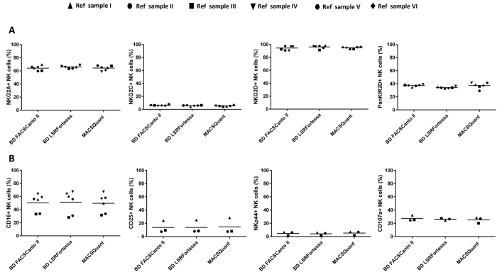
Figure 3. Evaluation of NK cell phenotypes and function marker expression between different flow cytometers.
Expression levels for drop-in antigens in the NK cell phenotype (A) and function panel (B) were analysed and compared across different centers using the same reference samples (n = 6). NKG2A, NKG2C, NKG2D, KIR2D and CD16 expression in BD LSRFortessaTM, BD FACSCantoTM II and MACSQuant® Analyzer 10 were measured for NK phenotype panel (A). Similarly, for NK cell function panel analysis, three time points of reference sample PBMC were stimulated with A431 cells and their percentage of NK cells positive for CD107a, CD25 and NKp44 levels were measured (B). Statistical analysis was performed using the non-parametric Kruskal-Wallis test.

