Fig. 5.
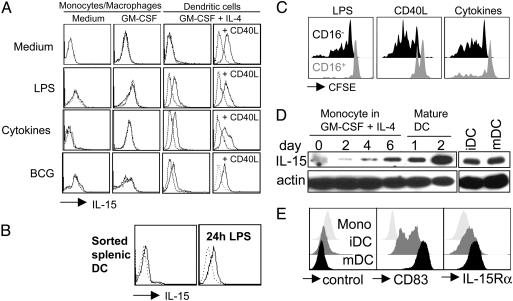
DCs present IL-15 on their surface upon maturation. DCs derived from monocytes by culture in the presence of GM-CSF and IL-4 (GM-CSF + IL-4) (A) and DCs directly isolated from spleen (B) present IL-15 on their surface upon maturation with LPS, proinflammatory cytokines (IL-1β, IL-6, TNF-α, and prostaglandin E2) or live bacillus Calmette–Guérin. Monocyte/macrophages cultured without cytokines (Medium) or in the presence of GM-CSF alone (GM-CSF) and then activated by the same stimuli used for DC maturation failed to express IL-15 on their surfaces. Addition of CD40L-transfected cells to DC cultures (CD40L) in the last 48 h was sufficient, even in the absence of inflammatory stimuli, to induce surface IL-15 expression in DCs but not in monocyte/macrophages (data not shown). Dotted lines represent isotype controls. One of three experiments is shown. (C) NK cells purified from peripheral blood were labeled with CFSE and cultured in the presence of autologous DCs matured by different stimuli. At day 6 of the cultures, cells were harvested, and CFSE dilution was evaluated (LPS, DCs matured by 100 ng/ml LPS; CD40L, DCs matured by CD40L-transfected cells; Cytokines, DCs matured by proinflammatory cytokines). One of three experiments is shown. (D) Western blot analysis of IL-15 at the indicated time points of DC differentiation from peripheral blood monocytes and with (mDC) and without (iDC) maturation by proinflammatory cytokines for 2 days. One of three experiments is shown. (E) Flow cytometric analysis of CD83 (Center) and IL-15Rα (Right) on monocytes (Mono), immature DCs (iDC; day 8) and mature DCs (mDC; day 8 and last 2 days incubated with proinflammatory cytokines). As a control for the polyclonal anti-IL-15Rα goat antibody a goat anti-mouse polyclonal antibody was used (control). One of three experiments is shown.

