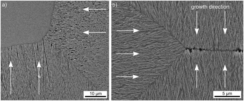
Figure 15.
SEM-micrographs of growth front collision zones in (a) the partially crystallized sample also featured in Fig. 13, and (b) a fully crystallized sample where no uncrystallized glass remains in the bulk. The main growth direction in the respective growth domains is indicated by arrows.

