Fig. 2.
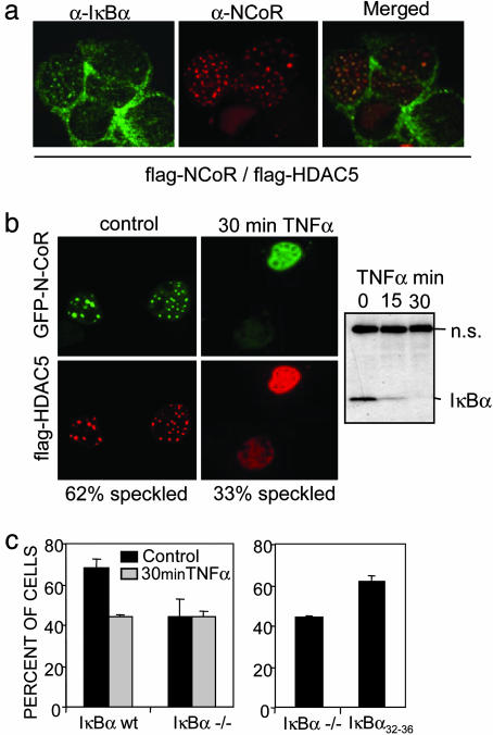
Endogenous IκBα colocalizes with N-CoR/HDAC5 nuclear speckles. (a) Colocalization of endogenous IκBα with ectopic N-CoR/HDAC-5-containing nuclear speckles [confocal image (×630)]. (b) Subcellular localization of GFP-N-CoR and flag-HDAC5 in cotransfected 293T cells before (Left) and after 30 min of TNF-α treatment (Right). A minimum of 200 cells were counted for each condition in an Olympus BX-60 microscope [confocal image (×630)]. The α-IκBα immunoblot shows TNF-α-induced IκBα degradation. (c) Percentage of cells displaying N-CoR/HDAC5 speckles in IκBα+/+ and IκBα–/– MEFs cotransfected with GFP-N-CoR and flag-HDAC5 in the control or after 30 min of TNF-α treatment (Left) or in the IκBα–/– MEFs transiently transfected with IκBα32–36 (Right). A minimum of 200 cells were counted for each condition in an Olympus BX-60 microscope.

