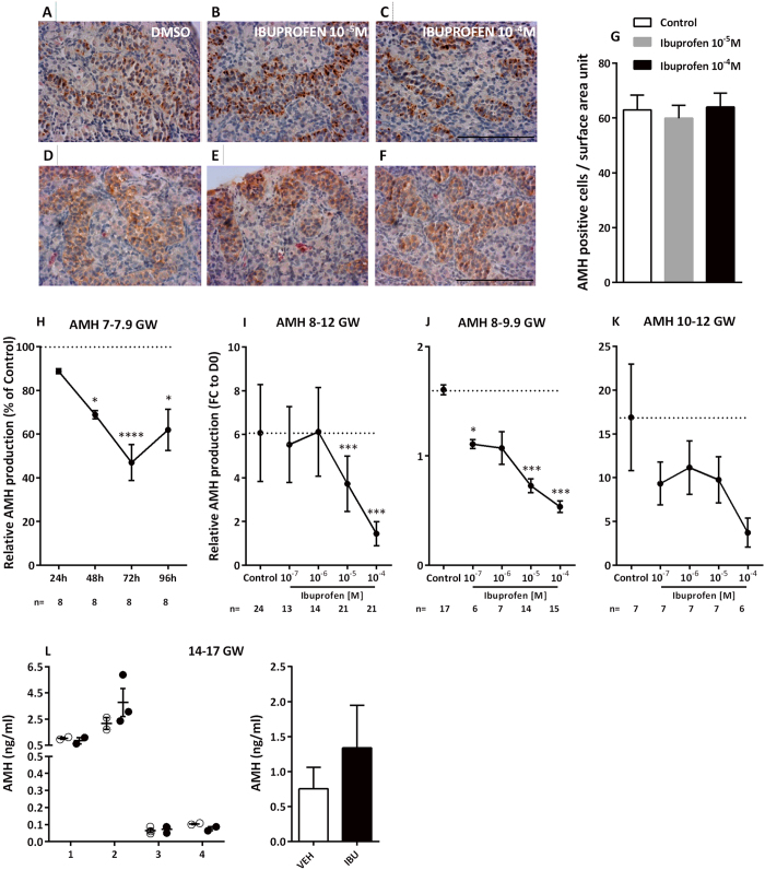
Figure 4. Ibuprofen and Sertoli cell function.
(A–F) Representative images of KRT18 (A–C) and AMH (D–F) immunostaining in cultured explants of 7–7.9-gestational week (GW) human fetal testis. KRT18 and AMH appear brown (3,3-diaminobenzidine tetrahydrochloride staining), and sections were counterstained with hematoxylin. Scale bar = 100 μm. (G) Sertoli cell numbers were determined by counting AMH-positive cells in control testes (white bars) and testes treated with 10−5 M (grey bars) and 10−4 M (black bars) of ibuprofen for 72 h. Data are presented as the number of cells per surface area unit (0.01 mm2) (means ± SEM) based on 1 or 2 explants per treatment in 7 fetuses. A Wilcoxon test was performed for pairwise comparisons. (H) AMH production after 24, 48, and 72 h of exposure to DMSO (Control) or 10−5 M of ibuprofen in 7–7.9 GW human fetal testes. Results are expressed as fold changes from the control testis (% of Ctrl). Values are means ± SEM for 7 testes from 7 fetuses. Repeated measures analysis of variance (ANOVA) on ranks was performed (*p < 0.05, ****p < 0.0001). (I–K) AMH production after culture of 8–12 GW (I); 8–9.9 GW (J); and 10–12 GW (K) human fetal testes in the presence of the control solvent DMSO (Control) or 10−7–10−4 M of ibuprofen. Results are expressed as fold change from the first day of culture (FC to D0). Values are means ± SEM of 6–17 testes from 6–17 fetuses for the 8–9.9 GW, and of 6–7 testes from 6–7 fetuses for the 10–12 GW. ANOVAs with a random fetus effect were performed using unstructured covariance matrices. A Wilcoxon test was performed for pairwise comparisons (*p < 0.05; ***p < 0.001). (L) Plasma AMH in individual host mice carrying human fetal testis xenografts (14–17 GW; n = 4 fetuses) after 7d exposure to vehicle (Corn Oil; open circles) or ibuprofen (10 mg/kg 3 times daily; closed circles) with overall mean ± SEM for vehicle (white bars) and ibuprofen (black bars). Data analyzed by two-way ANOVA.

