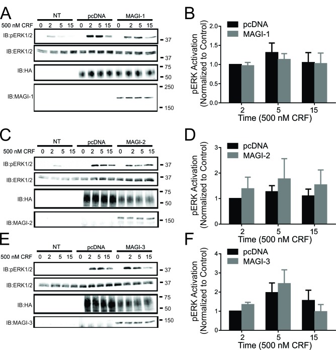
Figure 4.
Effect of MAGI proteins overexpression on CRFR1-mediated ERK1/2 signaling. (A) Representative immunoblot showing ERK1/2 phosphorylation in response to 500 nM CRF treatment for 0, 2, 5, and 15 min in non-transfected (NT) HEK293 cells, and HEK293 cells transfected with HA-CRFR1 along with either pcDNA or His-MAGI-1. Also shown are corresponding immunoblots for total ERK1/2, MAGI-1 and HA-CRFR1 protein expression. (B) Densitometric analysis of ERK1/2 phosphorylation in response to 500 nM CRF treatment for 0, 2, 5, and 15 min. The data represent the mean ± SEM of four independent experiments. (C) Representative immunoblot showing ERK1/2 phosphorylation in response to 500 nM CRF treatment for 0, 2, 5, and 15 min in non-transfected (NT) HEK293 cells, and HEK293 cells transfected with HA-CRFR1 along with either pcDNA or MAGI-2. Also shown are corresponding immunoblots for total ERK1/2, MAGI-2 and HA-CRFR1 protein expression. (D) Densitometric analysis of ERK1/2 phosphorylation in response to 500 nM CRF treatment for 0, 2, 5, and 15 min. The data represent the mean ± SEM of six independent experiments. (E) Representative immunoblot showing ERK1/2 phosphorylation in response to 500 nM CRF treatment for 0, 2, 5, and 15 min in non-transfected (NT) HEK293 cells, and HEK293 cells transfected with HA-CRFR1 along with either pcDNA or MAGI-3. Also shown are corresponding immunoblots for total ERK1/2, MAGI-3 and HA-CRFR1 protein expression. (F) Densitometric analysis of ERK1/2 phosphorylation in response to 500 nM CRF treatment for 0, 2, 5, and 15 min. The data represent the mean ± SEM of six independent experiments.

