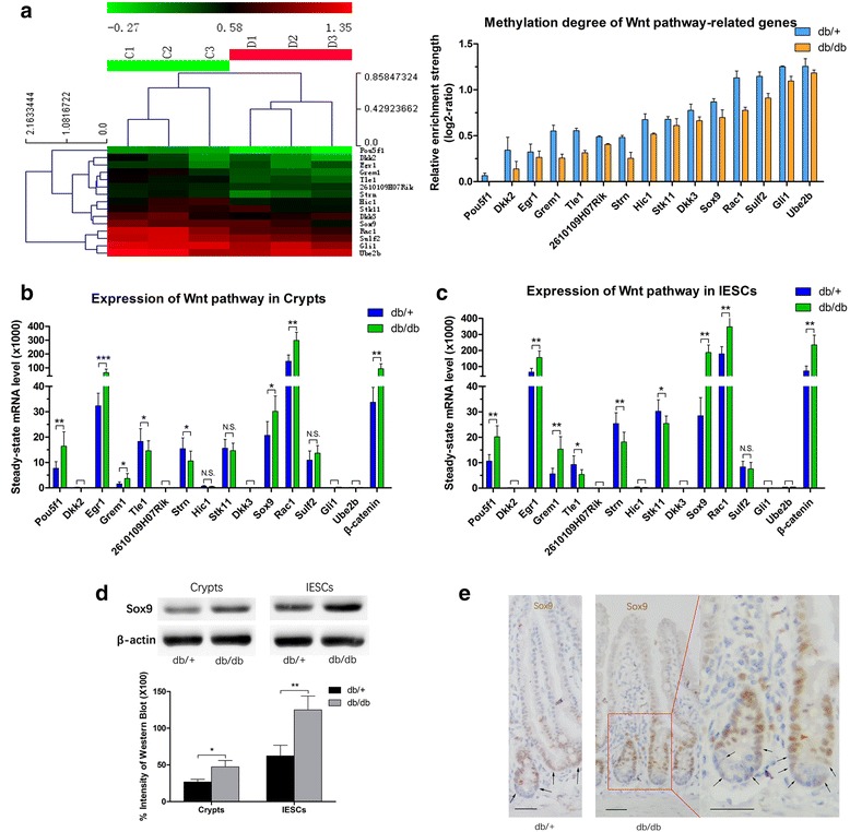
Fig. 3.

Promoter methylation levels and gene expression of Wnt signaling-related genes. a (left) Hierarchical clustering analysis on the basis of the DMEP-related genes related with Wnt signaling. Each row indicates a DMEP-related gene, and the corresponding gene name is indicated on the right. Each column indicates a sample analyzed. Methylation levels range from unmethylated (green) to fully methylated (red), as indicated by the color legend at the top of the graph. a (right) The degree of methylation of Wnt signaling pathway-related genes among DMEP-related genes. The bar height reflects the degree of methylation. b, c The mRNA levels of Wnt signaling-related genes in crypts and intestinal epithelium stem cells (IESCs) using the FACS cell population. d (upper) Western blot analysis of Sox9 using the MACS cell population, and d (lower) quantification of immunoblot bands from three repetitions of western blot experiments. e Immunohistochemical staining of Sox9 in the small intestinal epithelium. The arrows indicate cells expressing low levels of Sox9, which were considered IESCs. Scale bars = 50 μm. mRNA and protein levels are expressed relative to β-actin. Mean ± SE; n ≥ 6 (mRNA); n ≥ 3 (protein); *P < 0.05, **P < 0.01, ***P < 0.001 by t test. N.S. not significant
