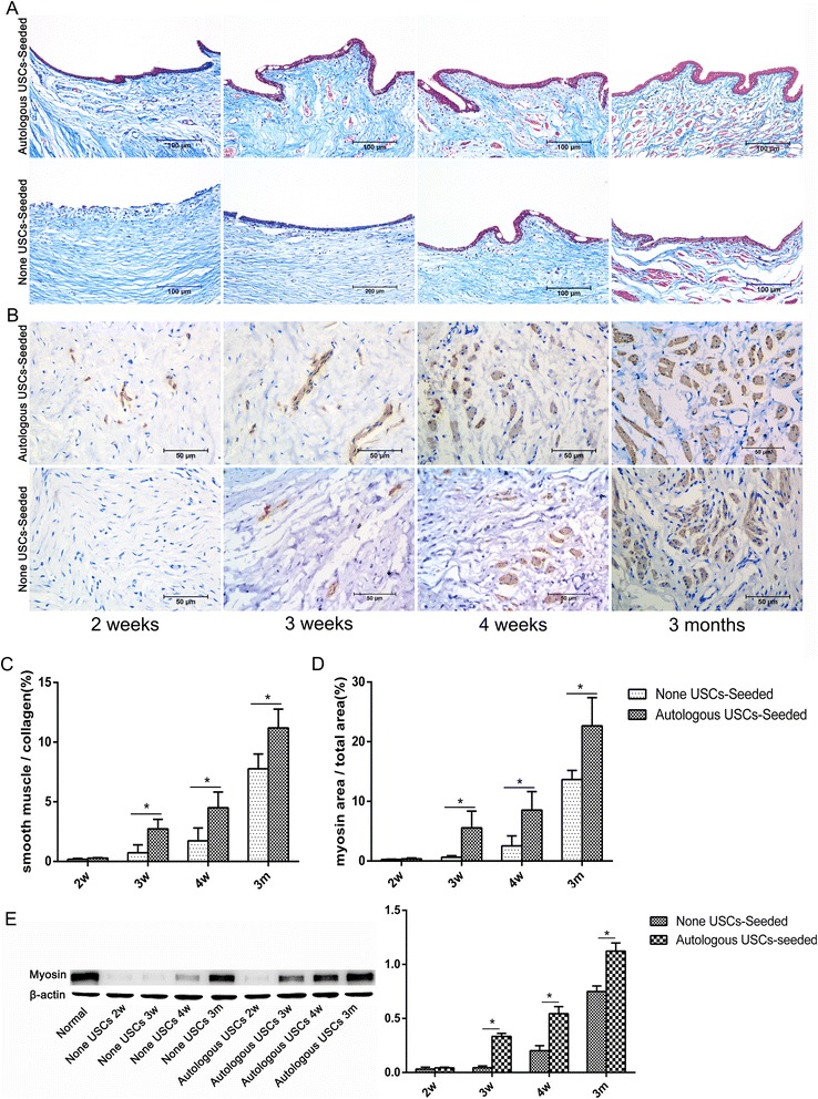
Fig. 8.

Histopathological evaluation of smooth muscle regeneration. a,b Smooth muscle regeneration. Representative Masson’s trichrome (a) and myosin IHC (b) staining in retrieved urethras 2, 3, and 4 weeks and 3 months after transplantation. In the 5% PAA-treated SIS only group, positive expression of myosin beneath the urothelium was observed 3 weeks after the surgery, and the smooth muscle content continued to increase over time. By contrast, expression appeared at 2 weeks in the autologous USC-seeded SIS group. Scale bar = 200 μm. c,d Image analysis. The smooth muscle-to-collagen ratio (c) assessed using Masson’s trichrome staining and the smooth muscle-to-total area ratio (d) assessed using myosin IHC staining at 3 and 4 weeks and 3 months in the two groups. These parameters were significantly higher in the autologous USC-seeded 5% PAA-treated SIS group than in the 5% PAA-treated SIS only group (*P < 0.05) at 3 and 4 weeks and 3 months. e Detection of SMC-specific markers (myosin) by Western blotting. m months, USC urine-derived stem cell, w weeks
