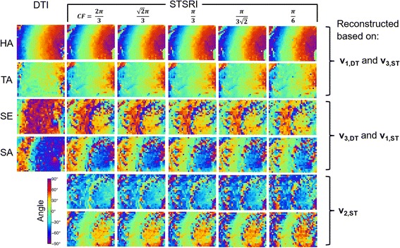
Fig. 8.

Helix angle (HA), transverse angle (TA), sheetlet elevation (SE) and sheetlet azimuth (SA) in LV myocardium reconstructed with DTI and STSRI using different reconstruction parameters. Here, high-resolution STSRI data with isotropic pixel size of 1.1 μm were analysed using centre frequencies (CF) of and corresponding kernel sizes of 73, 73, 93, 113, 173 voxels. The STSRI image corresponding to this region is given in Fig. 9
