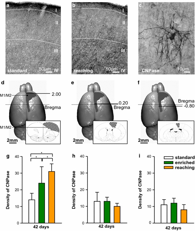
Fig. 4.

a, b Peroxidase-stained sections demonstrating CNPase+ oligodendrocytes after standard housing (a) compared to reaching training (b). c Higher magnification of CNPase+ cells. d–f Location of the different evaluated regions (Bregma 2.00, 0.20, −0.80) from the motor cortex (M1/M2) marked on the brain surface. The schematic detailed picture shows areas analysed [39]. g–i Quantification of the optical density of CNPase+ cells at Bregma 2.00 (g), Bregma 0.20 (h) and −0.80 (i). Significant differences are observed at Bregma 2.00 between the different activity groups compared to standard housing. Bars represent mean ± SD. Asterisks indicate significant differences (P < 0.05). Scale bars represent a 50 µm and b, c 10 µm
