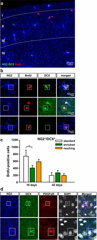
Fig. 6.

Immunocytochemical quantification of newly differentiated NG2+DCX+ subpopulation in the sensorimotor cortex of the different experimental groups. a Overview of proliferating BrdU+ (red) NG2+ (blue) DCX+ (green) cells at day 10 of the experiment. b Confocal images of triple-labelled sections with antibodies against BrdU (red) NG2 (blue) DCX (green) in the sensorimotor cortex. c Quantification of BrdU+NG2+DCX+ cells in the different experimental groups at days 10 and 42. d Confocal images of quadruple-labelled sections with antibodies against NG2 (blue), DCX (green), PDGFαR (red) and DAPI (white). NG2+/DCX+ cells co-labelled with an antibody against the PDGFα receptor in the adult cerebral cortex. Error bars represent S.D. Significant differences (P ≤ 0.05) are indicated by an asterisk. Scale bars a 50 µm and b 10 µm
