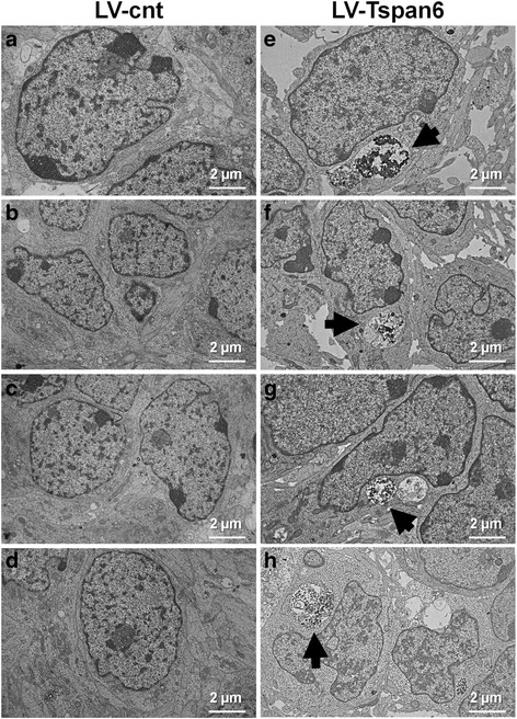
Fig. 10.

6 DIV primary cortical neurons derived from Tspan6 wt (Tspan6 +/Y) E14.5 embryos were infected with an empty lentiviral vector (LV-cnt) or a lentiviral vector overexpressing Tspan6 (LV-Tspan6). At 9 DIV neurons were washed and processed for EM analysis. Tspan6 wt neurons that overexpressed Tspan6 (e-h) show alterations by EM consisting of accumulation of electron dense material inside large vesicles (arrow) in the soma compared to control neurons (a-d)
