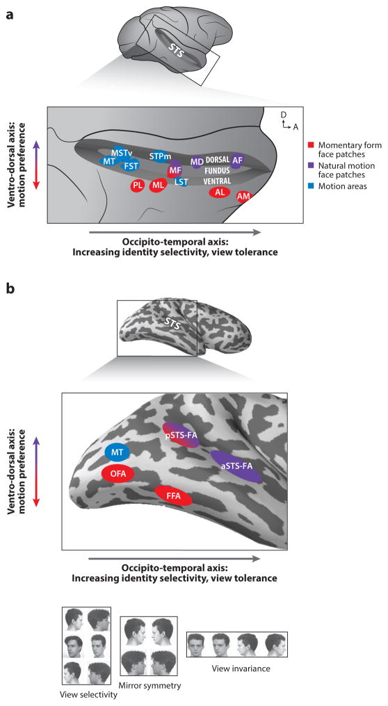
Fig. 1. Organization of Face Processing Systems in the Macaque and Human Brain.
a. Face patches in the macaque superior temporal sulcus (STS), shown here (top) opened on a lateral view of the brain, are functionally organized along an occipito-temporal (posterior-anterior) axis and a ventro-dorsal axis (expanded view, bottom). Arrows in upper right corner indicate dorsal (D) and anterior (A) direction. General motion areas (blue) and face areas (red and purple) are shown. Ventral areas (in the ventral bank of the STS and further ventrally) are selective for momentary form of faces (red), while areas at more medio-dorsal locations in the STS (fundus and dorsal bank) are selective for natural facial motion (purple). Hierarchical processing from view selective to view invariant representation is manifested in the posterior-anterior axis: representations are transformed from view-specific into increasingly view tolerant and facial identity-selective ones.
b. Face and motion areas (colors as in a) in the human brain are found ventrally in the lateral occipital cortex and the fusiform gyrus and dorsally in the superior temporal sulcus. A functional organization of hierarchical processing along the posterior-anterior axis and sensitivity to motion along the ventral-dorsal axis is found, similar to the one in the macaque brain. The ventral areas (red) show no sensitivity to motion whereas face areas in the STS are highly sensitive to moving faces. As in the macaque brain, the transformation along the occipito-temporal axis progresses via an intermediate representation that does not differentiate between left and right profile views (bottom). View selectivity was found in the lateral occipital cortex and mirror symmetry in the fusiform gyrus and posterior superior temporal sulcus.

