Fig. 3.
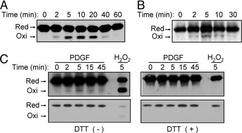
Detection of oxidized PTEN on the basis of an electrophoretic mobility shift in cells treated with exogenous H2O2 or with growth factors. (A) HeLa cells were incubated for the indicated times with 0.2 mM H2O2, after which cell lysates were subjected to alkylation with NEM and fractionated by nonreducing SDS/PAGE as described (11). Reduced (Red) and oxidized (Oxi) forms of PTEN were detected by immunoblot analysis with rabbit Abs to PTEN. (B) HeLa cells were incubated for the indicated times with 100 ng/ml EGF, after which cell lysates were subjected to alkylation with NEM followed by immunoprecipitation with a mAb (A2B1) to PTEN. The resulting precipitates were subjected to nonreducing SDS/PAGE and immunoblot analysis with rabbit Abs to PTEN as in A. (C) NIH 3T3 cells were transfected for 48 h with a mixture of Effectene (Qiagen) and a pCGN-derived vector encoding human PTEN tagged with HA at its NH2 terminus. The cells were subsequently stimulated for the indicated times with 25 ng/ml PDGF or for 5 min with 5 mM H2O2, after which cell lysates were analyzed as in B with the exception that the PTEN immunoprecipitates were either left untreated (Left) or treated with 1 mM DTT (Right) before immunoblot analysis with Abs to HA. The blots were exposed to x-ray film for 5 min (Upper) or 10 s (Lower).

