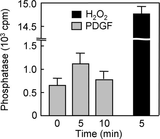
Fig. 5.
Detection by phosphatase assay of PTEN oxidized in NIH 3T3 cells stimulated with PDGF. Cells were incubated for 0, 5, or 10 min with 30 ng/ml PDGF or for 5 min with 5 mM H2O2, lysed in the presence of iodoacetic acid, and divided into three equal portions. PTEN was immunoprecipitated from each portion with a mAb to PTEN and then assayed for phosphatase activity under reducing conditions as described (15). Data are expressed as cpm of radioactivity released as Pi from the [32P]PIP3 substrate and are means ± SEM of the triplicate samples.

