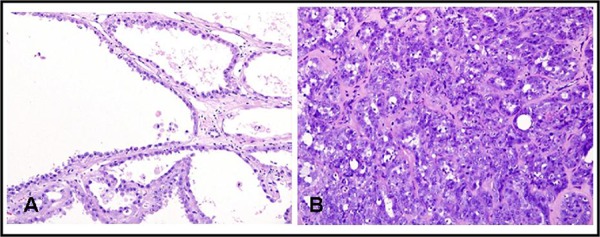
Figure 1.

Microscopic appearance of tubulocystic renal cell carcinoma. A, hematoxylin and eosin (H & E) staining shows tubulopapillary pattern with cystic spaces lined with cells having hobnail appearance (x200); B, H & E shows tubular and tubulopapillary structures in a hyalinized and fibrous stroma (x200).
