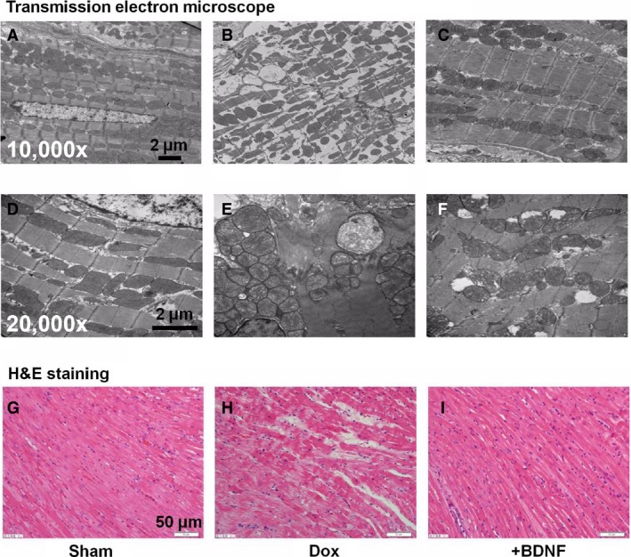
Figure 2.

Morphological alterations of LV myocytes in Dox and BDNF‐treated rats. (A–F) Transmission electron microscope images showed the ultrastructure changes of LV myocytes of sham, Dox and BDNF‐treated rats, at magnification 10,000× and 20,000×, respectively, scale bar: 2 μm. (G–I) Representative images of haematoxylin and eosin staining from left ventricles of sham, Dox and BDNF‐treated rats, at magnification 400×, scale bar: 50 μm.
