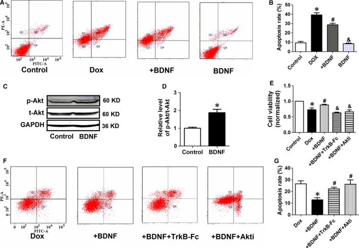
Figure 8.

BDNF inhibited Dox‐induced apoptosis of H9c2 cells by activating Akt. (A) Annexin V‐FITC/propidium iodide (PI) staining by flow cytometry was performed to detect the apoptosis of H9c2 cells. Early apoptosis (Annexin V‐FITC +/PI −, Q4), late apoptosis (Annexin V‐FITC +/PI +, Q2), and necrosis (Annexin V‐FITC −/PI +, Q1). (B) The quantitative presentation of apoptotic cell population by Annexin V‐FITC/PI staining. *P < 0.05 versus control, #P < 0.05 versus Dox, &P < 0.05 versus +BDNF, n = 3 each group. (C) Western blot analysis of the p‐Akt and t‐Akt. GAPDH served as the internal control. (D) Statistical results of p‐Akt/t‐Akt in H9c2 cells. *P < 0.05 versus control, n = 5 each group. (E) Effect of TrkB‐Fc and Akti on cell viability of BDNF‐treated H9c2 cells. *P < 0.05 versus control, #P < 0.05 versus Dox, and P < 0.05 versus +BDNF, n = 5 each group. (F) The quantitative presentation of apoptotic cell population by Annexin V‐FITC/PI staining. (G) Annexin V‐FITC/PI staining by flow cytometry was performed to detect the apoptosis. *P < 0.05 versus Dox, #P < 0.05 versus +BDNF, n = 3 each group.
