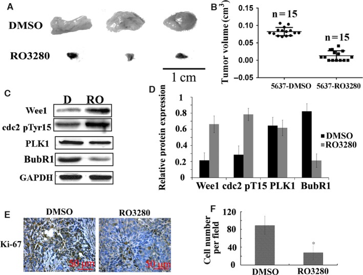
Figure 5.

RO3280 retards bladder cancer xenograft growth in vivo. (A) Representative images of tumours in DMSO‐treated 5637 cell‐transplanted mice (DMSO) and RO3280‐treated 5637 cell‐transplanted mice (RO3280). (B) The tumour volumes were measured at the indicated number of days after mice were transplanted with 5637‐DMSO and 5637‐RO3280 cells. The data are presented as the mean ± S.E.M. (C) Band intensities indicate Wee1 expression, CDC2 (Thr14/Tyr15) phosphorylation, PLK1 and BubR1 expression in 5637 xenograft with DMSO or RO3280 treatment. The data are presented as the mean ± S.E.M. (D) The ratio of the optical density of target protein and GAPDH of the same sample using Western blotting was calculated and expressed graphically (*P < 0.05, the data are presented as the mean ± S.E.M.). (E) Cell proliferation was evaluated using Ki‐67 immunohistochemistry in xenografts. (F) Statistical analysis of Ki‐67‐positive cells from C. The data are presented as the mean ± S.E.M. *P < 0.05.
