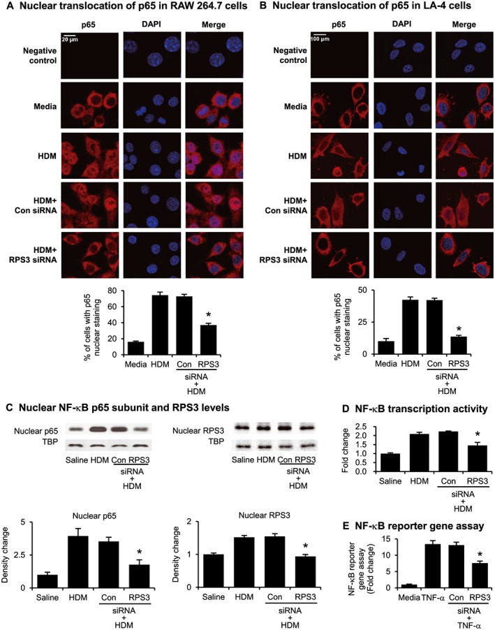
Figure 7.

Effects of RPS3 gene silencing on NF‐κB translocation and activity. Nuclear translocation of p65 induced by HDM was captured using immunofluorescence staining in (A) RAW 264.7 and (B) LA‐4 cells. The percentage of cells with p65 nuclear staining was quantified. Negative control panels were probed with the secondary antibody only to indicate potential background or non‐specific staining. (C) Immunoblot of nuclear NF‐κB subunit p65 and RPS3 accumulation. Mouse lung nuclear proteins were separated by 10% SDS‐PAGE and probed with anti‐p65, anti‐RPS3 or anti‐TATA binding protein (TBP) mAb. TBP were used as a nuclear protein loading control (n = 7–9). (D) Nuclear p65 DNA‐binding activity was determined using a TransAM™ p65 transcription factor elisa kit (n = 7–9). (E) NF‐κB reporter gene assay in NF‐κB luciferase reporter NIH/3T3 stable cell line pretreated with RPS3 siRNA and then stimulated with TNF‐α. Results are expressed as fold change relative to media control. The luciferase assay was conducted in duplicate wells for each sample, with three independent experiments. Values are shown as means ± SEM. *Significant difference from control siRNA, P < 0.05.
