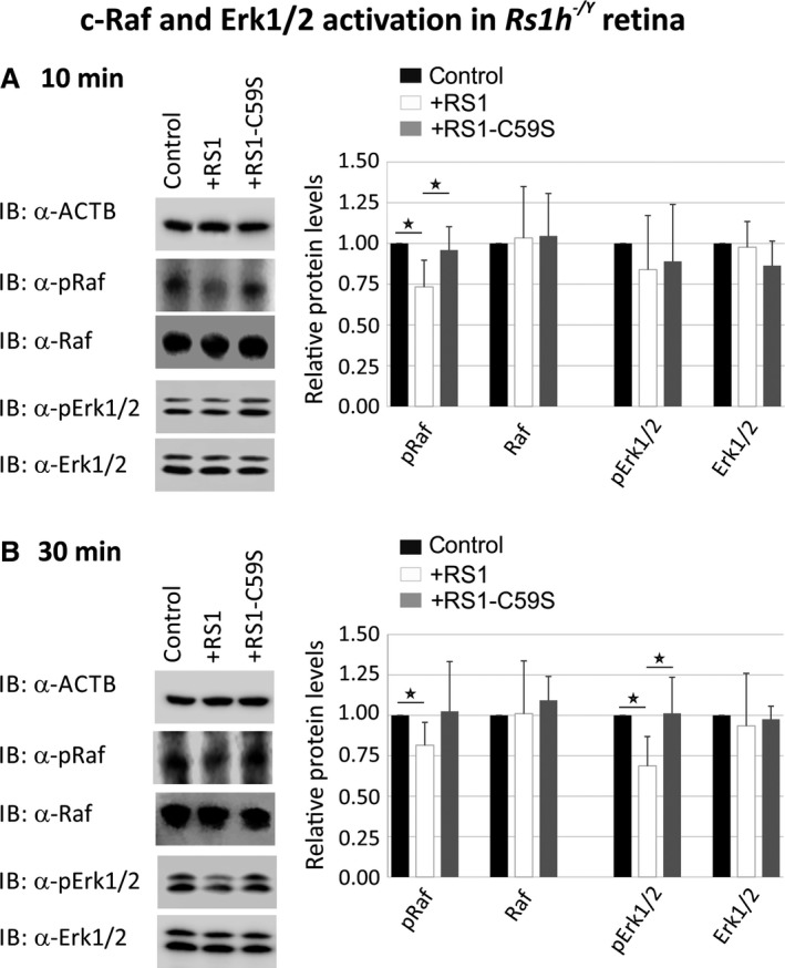
Figure 4.

Influence of retinoschisin on the ERK pathway in murine Rs1h −/Y retinal explants. Retinoschisin‐dependent c‐Raf and Erk1/2 phosphorylation in murine Rs1h −/Y retinal explants. Retinal explants harvested 10 days after birth (P10) were treated for 10 min. (A) or 30 min. (B) with retinoschisin, RS1‐C59S or control protein (purified from supernatant of empty expression vector‐transfected cells). Subsequently, the retinal lysates were subjected to Western blot analyses with antibodies against phosphorylated c‐Raf (pRaf), total c‐Raf (Raf), phosphorylated Erk1 and Erk2 (pErk1/2), total Erk1 and Erk2 (Erk1/2), as well as ActB as a control. (A and B) Densitometric quantification was performed with immunoblots from five independent experiments. Signals for pRaf Raf, pErk1/2 and Erk1/2 were normalized against ActB and calibrated against the control. Data represent the mean + S.D. Asterisks mark statistically significant differences (*P < 0.05).
