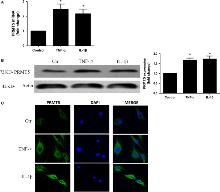
Figure 2.

Up‐regulated the expression of PRMT5 by proinflammatory mediators in RA FLSs. Cultured RA FLSs were stimulated with 10 ng/ml TNF‐α or 10 ng/ml IL‐1β. (A) Expression of PRMT5 mRNA was analysed by qPCR. Data were normalized with the GAPDH gene as control. (B) Expression of PRMT5 in the protein level was measured by Western blotting. Data were presented after normalization by β‐actin (right panel). (C) IF staining of anti‐PRMT5 antibody (green) in RA FLSs. The nuclei of cells were counterstained with DAPI (blue). Original magnification 630×. The pictures represented three experiments. Data shown are representative of five independent experiments from three different RA patients. *P < 0.05 versus control.
