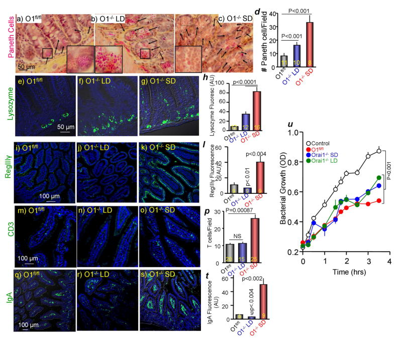
Figure 5. Analysis of intestinal innate immunity in Orai1fl/fl and Orai1−/− mice.
(a–d) Phloxine B/tartrazine staining of intestinal sections obtained from Orai1fl/fl (a) and Orai1−/− mice maintained on liquid (b) or solid diet (c). (d) Shows the average number of Paneth cells/field analyzed in the indicated number of fields obtained from 3 mice in each line.
(e–h) Intestinal sections from 3 Orai1fl/fl, 3 Orai1−/− mice on liquid diet and 4 Orai1−/− mice on solid diet were stained for lysozyme (green) and counterstained for DAPI (blue). (h) Shows the average lysozyme fluorescence in the indicated number of fields.
(i–l) Intestinal section from the indicated mice as in (e–h) were stained for RegIIIγ (green) and counterstained for DAPI (blue). (l) Shows the average RegIIIγ fluorescence.
(m–p) Intestinal section from the indicated mice as in (e–h) were stained for CD3 T cells (green) and counterstained for DAPI (blue). (p) Shows the average number of T cells/field analyzed in the indicated number of fields.
(q–t) Intestinal section from the indicated mice as in (e–h) were stained for IgA (green) and counterstained for DAPI (blue). (t) Shows the average IgA fluorescence in the indicated number of fields.
(u) Secreted duodenal antibacterial killing activity of Orai1fl/fl (red) and Orai1−/− mice maintained on liquid (green) or solid diet (blue). Results are mean±s.e.m of extracts obtained from 3 mice in each line. Deletion of pancreatic acinar Orai1 did not inhibit intestinal antibacterial secretion and killing.

