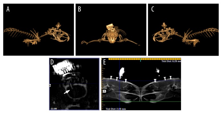
Figure 1.
Localization of implanted microwire array is confirmed in the rACC using three-dimensional reconstructions and radiographs from CBCT. (A) Right lateral view. (B) Anterior-posterior view. (C) Left lateral view. (D) The cross-section of the rACC showing implant placement (arrow). (E) Reconstructed panoramic radiograph. rACC – rostral anterior cingulate cortex; CBCT – cone-beam computed tomography.

