
Figure 10.
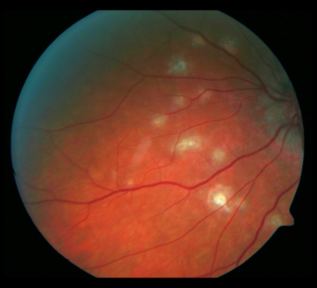
Whitish, irregularly shaped foci visible on the nasal side of the optic disc. The fundus of the left eye 18 months later. Progression of the macular fibrosis and the foci on the nasal side of the optic disc.
Official websites use .gov
A
.gov website belongs to an official
government organization in the United States.
Secure .gov websites use HTTPS
A lock (
) or https:// means you've safely
connected to the .gov website. Share sensitive
information only on official, secure websites.
Whitish, irregularly shaped foci visible on the nasal side of the optic disc. The fundus of the left eye 18 months later. Progression of the macular fibrosis and the foci on the nasal side of the optic disc.