Figure 8.
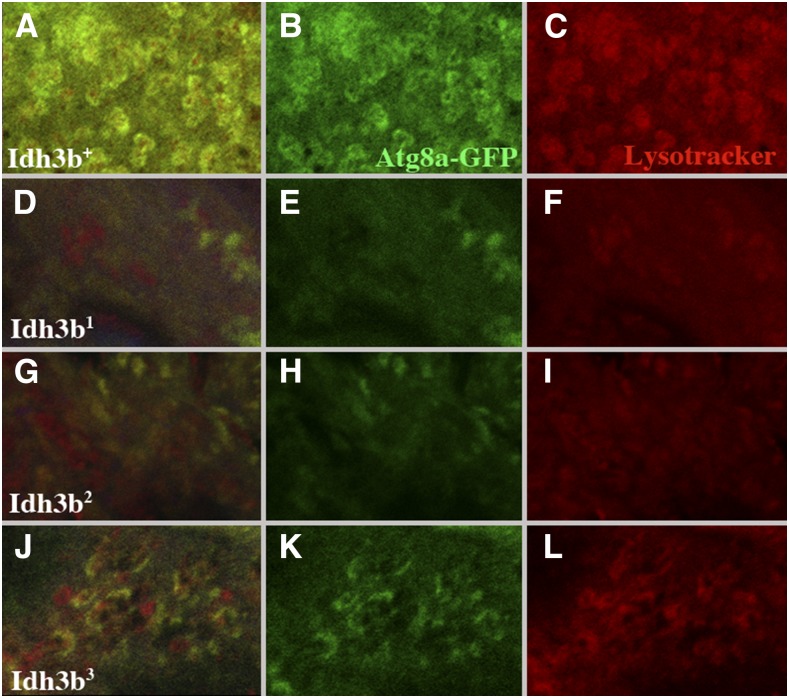
Autophagy is blocked in Idh3b-mutant salivary gland cells. All images show salivary gland cells from 8 to 12 APF prepupae, in which expression of UAS-Atg8a-GFP is driven by Sg-Gal4. Atg8a-GFP is shown in green, and LysoTracker staining is in red. (A–C) In Idh3b+/Df(3R)Exel6188 cells, Atg8a-GFP labeled vesicles are densely packed, and these vesicles colabel with LysoTracker, identifying them as autolysosomes. (D–I) In Idh3b1/Df(3R)Exel6188 and Idh3b2/Df(3R)Exel6188 cells, Atg8a-GFP-labeled vesicles are dramatically reduced in number, and LysoTracker staining is much reduced, indicating an almost complete block in autophagy. (J–L) Idh3b3/Df(3R)Exel6188 cells show an intermediate phenotype, in which Atg8a-GFP vesicles are partially formed, and LysoTracker staining is weakened, and incompletely associated with Atg8a-GFP fluorescence.

