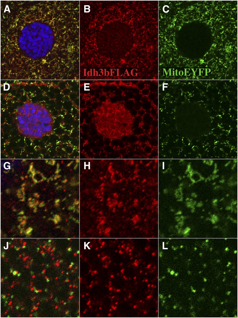
Figure 9.
Expulsion of Idh3b from mitochondria after fragmentation. All images are from larvae of the genotype UAS-HA/FLAG-Idh3b/+; tub-Gal4/mito-EYFP. (A–C) Salivary gland cell from a mid third-instar larva. FLAG-Idh3b and mito-EYFP colocalize. The nucleus (marked by DAPI staining in blue) shows no mito-EYFP fluorescence, and is very weakly labeled for FLAG-Idh3b. (D–F) Salivary gland cell from a late (wandering) third-instar larva. Mitochondrial fragmentation has taken place. FLAG-Idh3b staining and mito-EYFP are no longer coincident (see merged image in D). Note that FLAG-Idh3b staining within the nucleus has become more intense (E), but that mito-EYFP staining continues to be excluded from the nucleus (F). The dark lacunae in these images are secretory vacuoles, which are prominent at this time. (G–I) High magnification of part of a mid third-instar salivary gland cell. Note that FLAG-Idh3b staining (H) and mito-EYFP (I) largely colocalize (G). (J–L) High magnification of part of a late third-instar salivary gland cell. FLAG-Idh3b puncta (K) and mito-EYFP (L) are now almost mutually exclusive (merged image in J).

