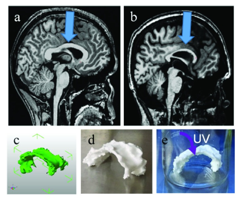
Figure 1. 3D models and prints.
( a– b) T1-weighted brain MRI with resolution of 1×1×1 mm, midsagittal slice, arrow pointing at corpus callous in ( a) healthy control and ( b) MPS I subject. ( c) CAD image of MPS I corpus callosum taken at five adjacent slices in each hemisphere. ( d) 3D printed MPS I corpus callosum with poly(lactic-acid) on a Makerbot Replicator 5X. ( e) UV sterilizing the print overnight.

