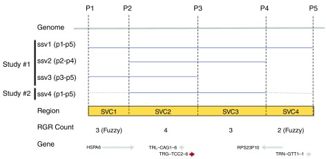
Figure 1. An alignment of variants ssv1-ssv3 (blue lines) with the genome (grey line) between positions P1 and P5.
Reference genomic regions SVC1-SVC4 (yellow box) are demarcated by overlap and non-overlapping positions (P1-P2, P2-P3, etc.) between SSVs. The observed SVC counts and the genes are shown on the bottom.

