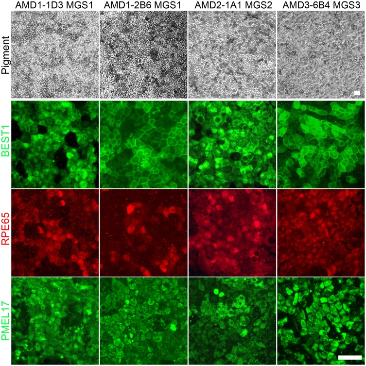
Fig 4. Immunohistochemistry of iPSC-derived RPE cell lines from donors with and without AMD.
RPE cell lines generated from iPSC lines derived from donors with and without AMD were expanded in culture. Phase microscopy images were taken to illustrate the pigmented monolayer. Immunohistochemistry and fluorescent microcopy was used to detect the expression pattern of characteristic protein markers of RPE, the ion channel BESTROPHIN, 65kDa Retinal Pigment Epithelium-Specific Protein RPE65 and the pre-melanosome protein PMEL17. (Scale bar 50 μm).

