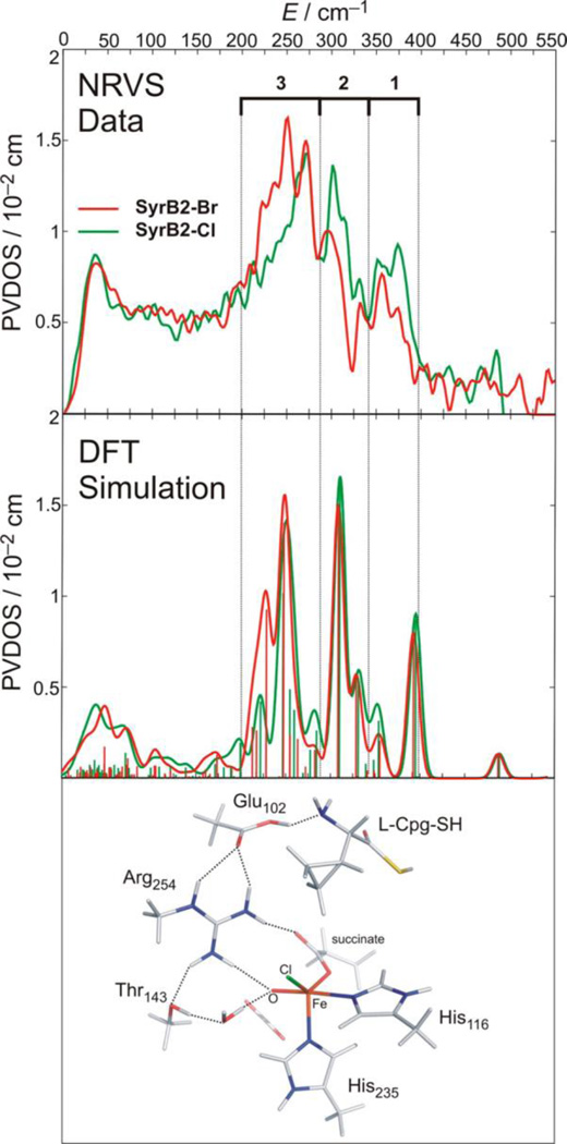
Figure 7.
Top: NRVS spectra for SyrB2-Cl (green) and SyrB2-Br (red). Middle: DFT NRVS simulations derived from the structure that best reproduces the experimental data. Bottom: Experimentally derived structure of the FeIV=O intermediate of SyrB2, which has a 5C trigonal bipyramidal geometry and an ~C3 axis. Adapted from ref 69.

