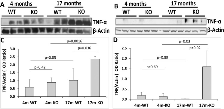
Fig 9. Increased protein levels of TNF-α in the RPE/choroid and retina of aged Cxcr5-/- mice.
(A and B) Western blots (WB) of TNF-α and β-actin with the RPE/choroid (A) and retina (B). The RPE/Choroid and retinal protein samples from three individual mice were used for each group. Protein blot was first probed by anti-TNF-α antibody. After stripping and washing, the same blot was re-probed by anti-β-actin antibody. (C and D) WB quantification of RPE/choroid (C) and retina (D). The results were the mean optical density (OD) ratio of TNF-α and β-actin (± SD; n = 3). 4/17m-WT = 4/17-month-old C57BL/6 wild type mice. 4/17m-KO = 4-month-old Cxcr5 knockout mice.

