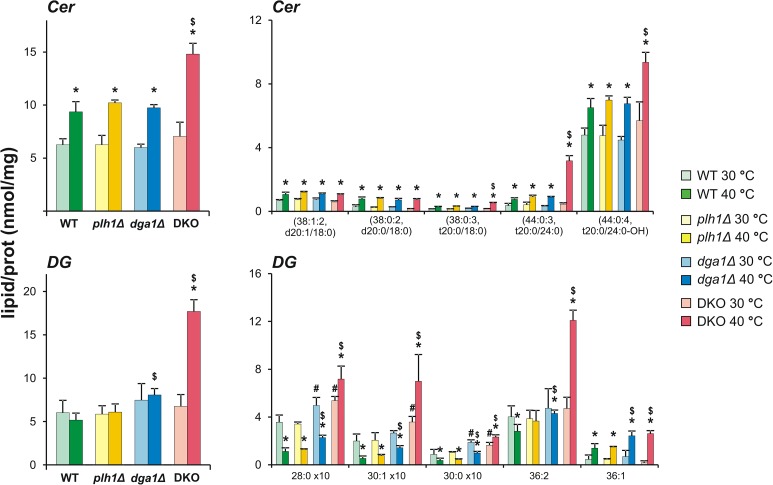
Fig 5. Growth arrest of heat-stressed DKO cells correlates with enhanced signalling lipid generation.
Changes in the amounts of Cer and DG by lipid class and species levels are shown. Cells were untreated (30°C) or stressed at 40°C for 1 h. Values are expressed as mean ± SD of lipid/protein values (nmol/mg), n = 3 for plh1Δ and dga1Δ, n = 4 for DKO, and n = 7 for WT; * p<0.05 (30°C vs 40°C), # p<0.05 (WT vs mutants at 30°C), $ p<0.05 (WT vs mutants at 40°C).

