
FIGURE 4.
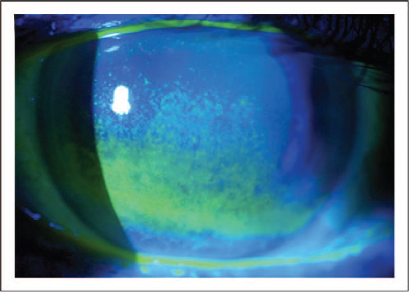
Corneal fluorescein staining. Image from a patient with aqueous deficiency. The disruption in the integrity of the corneal epithelium is highlighted by the application of fluorescein staining to the ocular surface. Moderate-to-severe staining with fluorescein is depicted in the photograph. Image courtesy of Karl Stonecipher, MD.