
FIGURE 8.
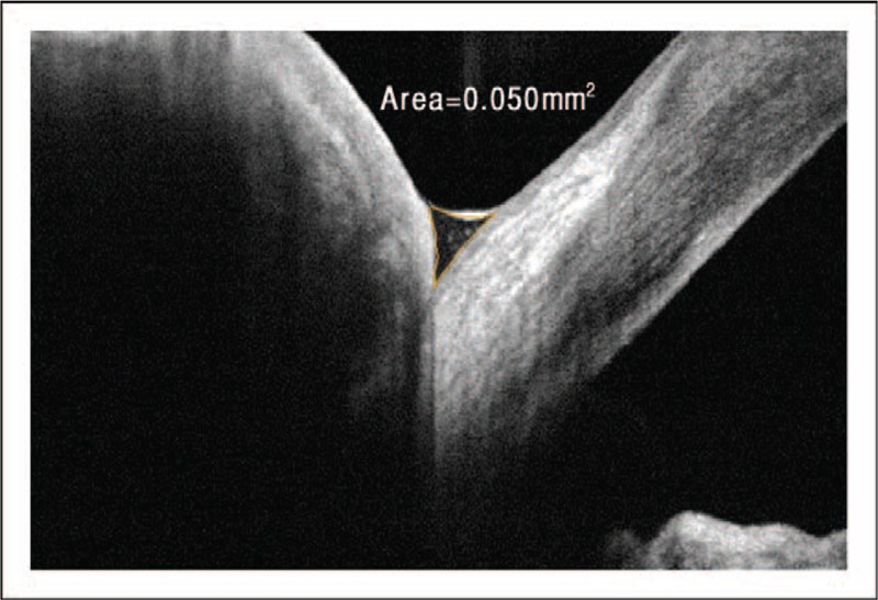
Evaluation of the tear film meniscus with optical coherence tomography (OCT). Image of the cross-sectional area of the tear film meniscus with OCT. Evaluation of the tear film with a high-resolution OCT allows for the characteristics of the tear film to be quantified. The area of the inferior meniscus was calculated to be 0.05 mm2 in this patient, which was within the normal range [77]. Image courtesy of Elizabeth Yeu, MD.