
FIGURE 9.
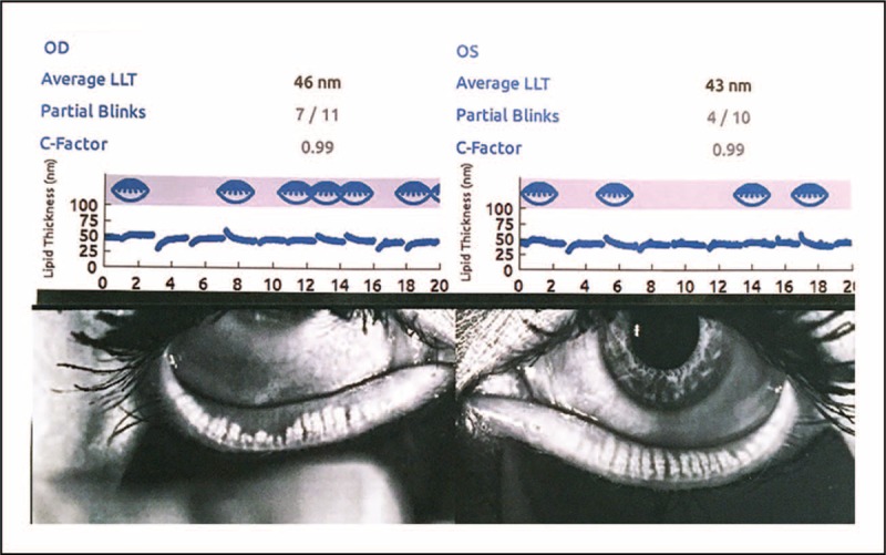
Visualization of the meibomian glands, tear film lipid layer, and partial blinking. Image of a patient with meibomian gland dysfunction (MGD) and exposure-related dysfunctional tear syndrome (DTS). Diagnostic systems designed for visualization of the meibomian gland (meibography) are also capable of providing an analysis of the thickness of the tear film lipid layer and an assessment of partial blinking. Note a decrease in lipid layer thickness of 46 nm in the right eye and 43 nm in the left eye; increased partial blinking OU; and meibomian gland truncation OD greater than OS. Image courtesy of Mark Milner, MD.