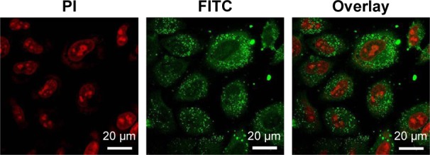
Figure 5.

Confocal laser scanning microscopy images of A875 cells after 2 h of incubation with C6-loaded CA-PLGA-TPGS NPs. C6-loaded NPs were green, and the cells were stained by PI (red). The cellular uptake was observed by overlaying images obtained by FITC channel (green) and PI channel (red). Magnification ×0.5 k.
Abbreviations: C6, coumarin-6; CA, cholic acid; PLGA, poly(lactide-co-glycolide); TPGS, D-α-tocopheryl polyethylene glycol 1000 succinate; NPs, nanoparticles; PI, propidium iodide; FITC, fluorescein isothiocyanate.
