Fig. 5.
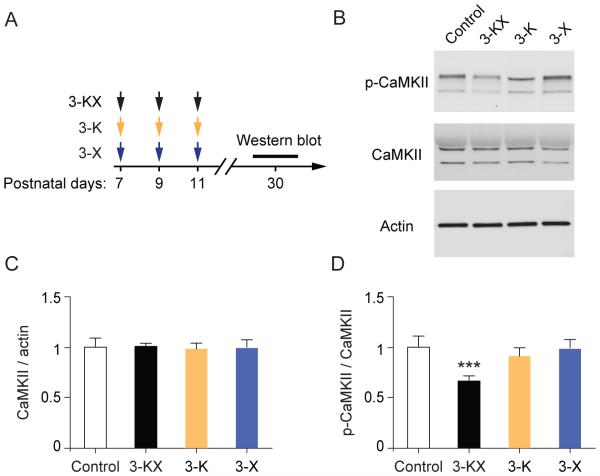
Multiple exposures to KX anesthesia at neonatal age result in impaired CaMKII signaling. (A) Timeline for antiesthetic exposure and Western blot analysis. (B) Synaptosome fractions were generated from the brains of 1-month-old mice, and probed with antibodies for CaMKII and p-CaMKII by Western blot. (C) Densitometric quantification of Western blots (n = 5 mice per group). The amount of total CaMKII is comparable between control and anesthetic groups. 3-KX vs. Control: t(8) = 0.1944, P = 0.8507; 3-K vs. Control: t(8) =0.3920, P =0.7053; 3-X vs. Control: t(8) =0.1649, P =0.8731; unpaired t test. (D) Phosphorylation levels of CaMKII were significantly decreased in mice with repeated exposure to KX anesthesia. 3-KX vs. Control: t(8) = 6.296, P = 0.0002, 3-K vs. Control: t(8) =1.429, P =0.1910; 3-X vs. Control: t(8) =0.2479, P =0.8105; unpaired t test. Data are presented as means ± SEM. ***P < 0.001.

