
Figure 8.
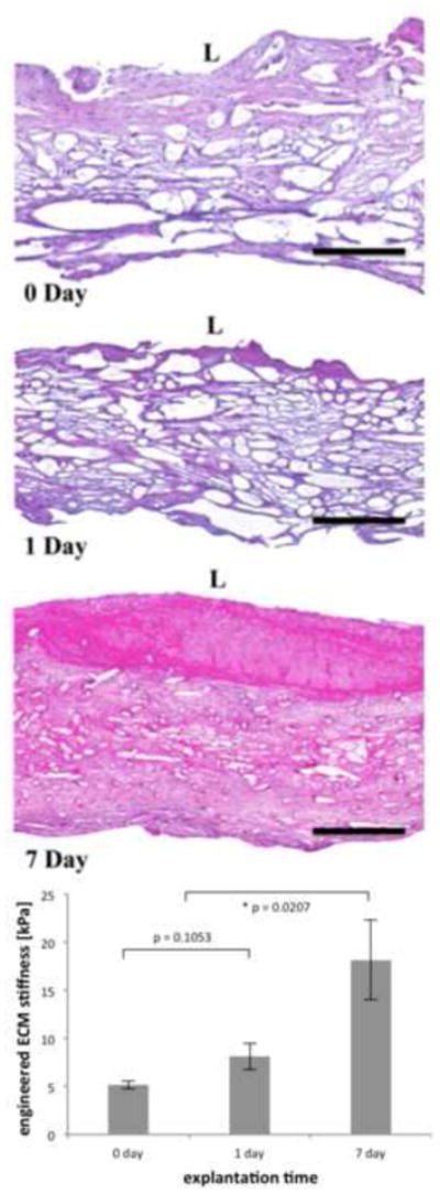
Representative transmural H&E histology of pulmonary valve conduits obtained post incubation (t = 0 day) and post-implantation in an ovine model (t = 1 and 7 day) of a parallel study [28, 29] (top) and ECM stiffness (bottom). Black scale bar represents 300 μm, L denotes the luminal, and collagen is stained in pale pink and cell nuclei in blue. The scaffold fibers are clearly noticeable at all time points. A substantial amount of void space is observed at the early time points, yet after 7 days of in vivo implantation, the engineered tissue was densely packed and a fair amount of inflammatory reaction on the lumen side is observed in this sample. ECM stiffness increased substantially after 7 days of in vivo implantation.