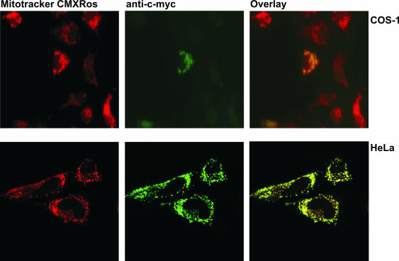
Figure 4.
Mitochondrial localization of hmtPAP in mammalian cells. COS-1 and HeLa cells were grown on coverslips and transiently transfected with pcDNA + hmtPAP plasmid, encoding c-myc-tagged hmtPAP. After incubation with the mitochondria-specific dye MitoTracker CMXRos (300 nM), fixation with 4% formaldehyde and permeabilization with 1% Triton X-100, the cells were immunostained with anti-c-myc monoclonal antibody, which was then visualized with FITC-conjugated antibody. Fluorescent images of MitoTracker (red) and c-myc-tagged hmtPAP (green) were taken by either a fluorescent (COS-1 cells) or a confocal (HeLa cells) microscope. Co-localization of hmtPAP and mitochondria appears yellow in digitally overlaid images.

