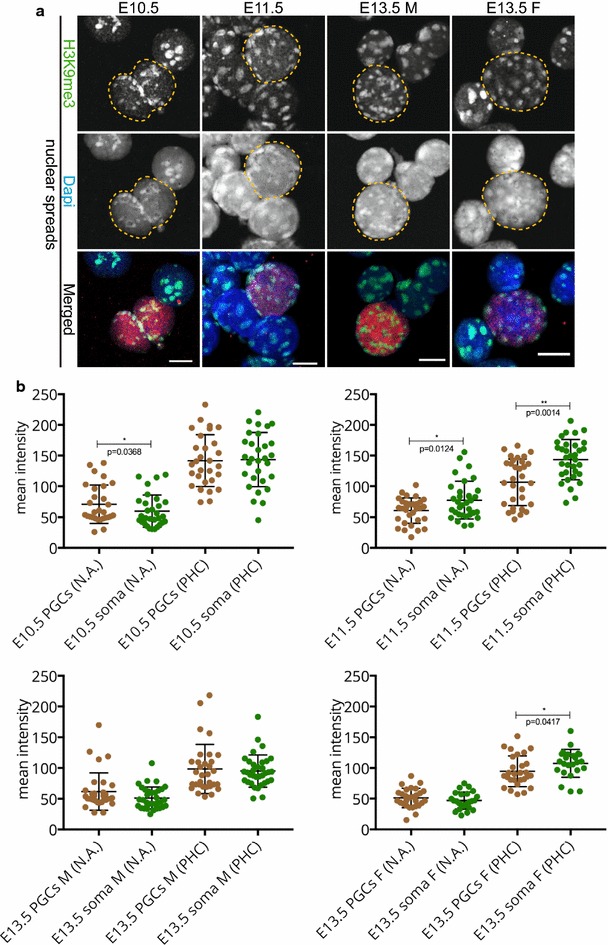
Fig. 2.

H3K9me3 signal enrichment persists on pericentric heterochromatin throughout germ cell development in spread preparations. a Spread preparations immunostained for H3K9me3 (green). E10.5 and E11.5 germ cells are marked with OCT4 (red), while E13.5 germ cells are marked with TRA98 (red). Representative germ cells are marked with yellow dashed circles. Scale bars represent 10 μm. b Quantification analysis of mean H3K9me3 levels in the whole nuclear area (N.A.) and pericentric heterochromatin regions (PHC) in the different embryonic stages examined. Four or more embryo trunks or gonads were pooled for the nuclear spread preparations and 20–30 PGC, and somatic nuclei were recorded. Asterisks (* or **) indicate significant differences
