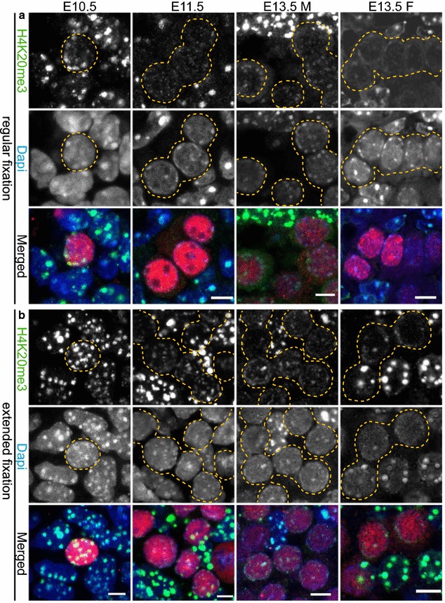
Fig. 6.

H4K20me3 enrichment is detected in pericentric heterochromatin of developing germ cells. a Paraffin sections of E10.5 and E11.5 embryo trunks and of E13.5 male and female gonads were immunostained using anti-H4K20me3 (green) after applying the regular fixation protocol. At E10.5 H4K20me3 is present in pericentric heterochromatin of PGCs and surrounding soma. At E11.5, H4K20me3 is lost from DAPI (blue)-dense regions of PGCs, while it is maintained in the somatic cells. At E13.5 H4K20me3 reappears in germ cells, but in substantially reduced levels compared to the surrounding gonadal somatic cells. b When applying the extended fixation protocol, H4K20me3 is retained at pericentric heterochromatin in PGC nuclei from E10.5 to E13.5. However, when compared to the H4K20me3 pattern in the surrounding somatic cells, the levels are reduced. E10.5 and E11.5 germ cells are marked with OCT4 (red), while E13.5 germ cells are marked with TRA98 (red). Using regular or extended fixation, two embryos or gonads were analysed per stage, and at least 20 PGC nuclei were recorded. Representative germ cells are marked with yellow dashed circles. Scale bars represent 5 μm
