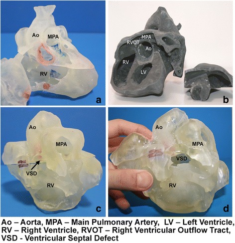
Fig. 1.

Respective physical models and features are as shown. a Normal heart: This model, created from Cardiac CT, is partitioned into 3 pieces, including an anterior portion (the right ventricular free wall) that can be removed to visualize the normal interventricular septum. The remaining superior and inferior portions can be separated to allow for visualization of the aorta and its position relative to the right ventricle. b Repaired tetralogy of Fallot heart from an adult: The model, created from Cardiac MRI is separated into 2 pieces, well fitted together via “Lego” peg depression. The cut in the main body allows for clear visualization of the pulmonary infundibular stenosis and overriding aorta. c Unrepaired tetralogy of Fallot heart from an infant: The 3D model, created from 3D echocardiogram, was partitioned into 2 pieces; a superior and inferior portion divided along the ventricular septal defect. d Unrepaired tetralogy of Fallot heart from an infant: Separating superior and inferior portions allows for clear visualization of the VSD as well as the aortic override relative to the VSD
