
Figure 7.
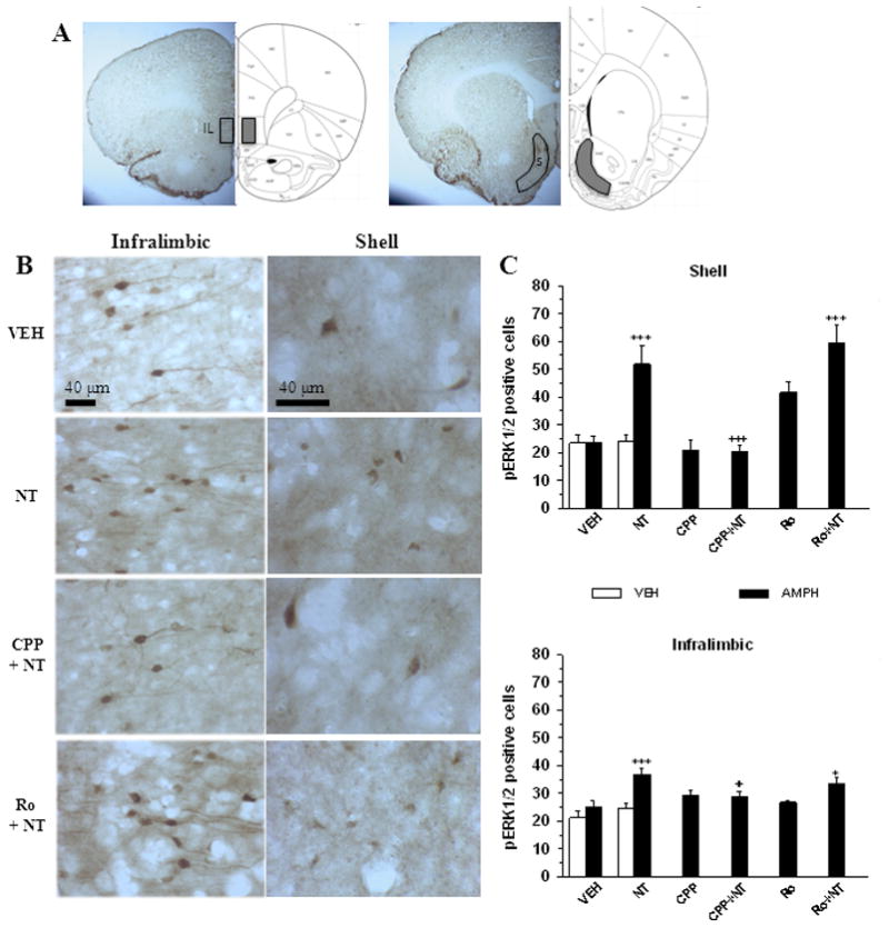
Amphetamine-induced pERK1/2 in the shell of the nucleus accumbens and the infralimbic cortex in animals pre-exposed to VM neurotensin. A. Representative coronal brain slices depicting the regions of infralimbic cortex (IL) and nucleus accumbens shell (S) where pERK1/2 immunoreactivity were sampled. B. Representative photomicrophaphs illustrating pERK1/2 immunoreactivity in the infralimbic cortex and shell of the nucleus accumbens in coronal brain slices from animals injected with systemic amphetamine (0.75 mg/kg, i.p.). Legend on the left indicates the treatment that each animal received during the training phase: Vehicle (VEH); D-Tyr[11]Neurotensin (NT); CPP + NT (CPP + NT); Ro04-5595 + NT (Ro + NT). C. Group means (± s.e.m) total number (shell) or total number per 0.2 mm2 (infralimbic) of pERK1/2 positive cells quantified in the shell (top panel) and the infralimbic cortex (bottom panel) after systemic vehicle (VEH, white) or amphetamine (AMPH, black) in different groups of animals that were injected during the training phase with VEH (n = 7 in each group), 1.5 nmol/side of D-Tyr[11]Neurotensin (NT; n = 7 in each group), 120 pmol/side of CPP with or without NT (CPP + NT and CPP, n = 6 in each group), or 1200 pmol/side of Ro04-5595 with or without NT (Ro + NT, n = 7; Ro, n = 6). Symbols indicate a statistically significant difference with VEH + AMPH group (*p < 0.05; ***p < 0.001) or with NT + AMPH (+p < 0.05; +++p < 0.001). See text for details.