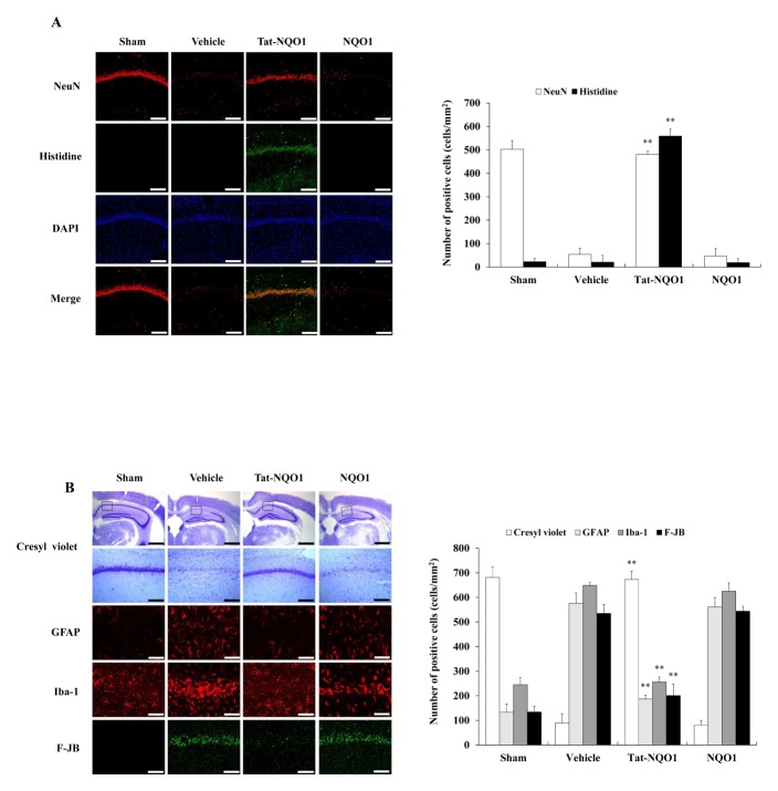
Fig. 4.
Effects of transduced Tat-NQO1 protein in an animal model of ischemia. Gerbils were treated with a single injection of Tat-NQO1 protein (2 mg/kg) before ischemia-reperfusion, and sacrificed after 7 days. Transduced Tat-NQO1 protein was analyzed by immunostaining, using anti-Histidine and DAPI staining. Ischemic neuronal damage was analyzed by NeuN-immunostaining (A). Neuronal cell viability was analyzed by Cresyl violet (CV), F-JB, Iba-1 and GFAP immunostaining. Relative numeric analysis of CV and F-JB, Iba-1, GFAP positive neurons in the CA1 region (B). Scale bar = 18.8 and 50 μm. **P < 0.01, significantly different from the vehicle group.

