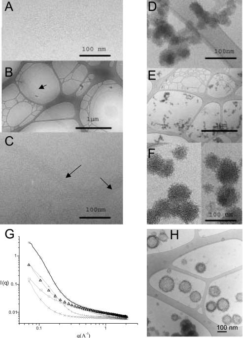
Figure 2.
Cryo-TEM micrographs and X-ray scattering of poloxamine 304 and poloxamine 304/DNA complexes in saline and in Tyrode's solution. (A) Cryo-TEM micrograph of a 10% poloxamine 304 in saline. (B) Cryo-TEM micrograph of 304/DNA complexes with 10% poloxamine 304 in saline. (C) Cryo-TEM micrograph of the same sample at higher magnification. (D) Cryo-TEM micrograph of poloxamine 304 in Tyrode's solution. (E) Cryo-TEM micrograph of a poloxamine 304/DNA complexes with 10% poloxamine 304 in Tyrode's solution. (F) The same sample at a higher magnification. (G) Small-angle X-ray scattering scans of: poloxamine 304 in saline (crosses), in Tyrode's solution (plus sign), of DNA in Tyrode's solution (open squares), of poloxamine 304/DNA complexes in saline (open triangles) and in Tyrode's solution (open circles). (H) Cryo-TEM micrograph of poloxamine 304/DNA complexes in Tyrode's solution interacting with unilamellar vesicles of phosphatidylcholine/phosphatidylglycerol (30:70, w:w).

