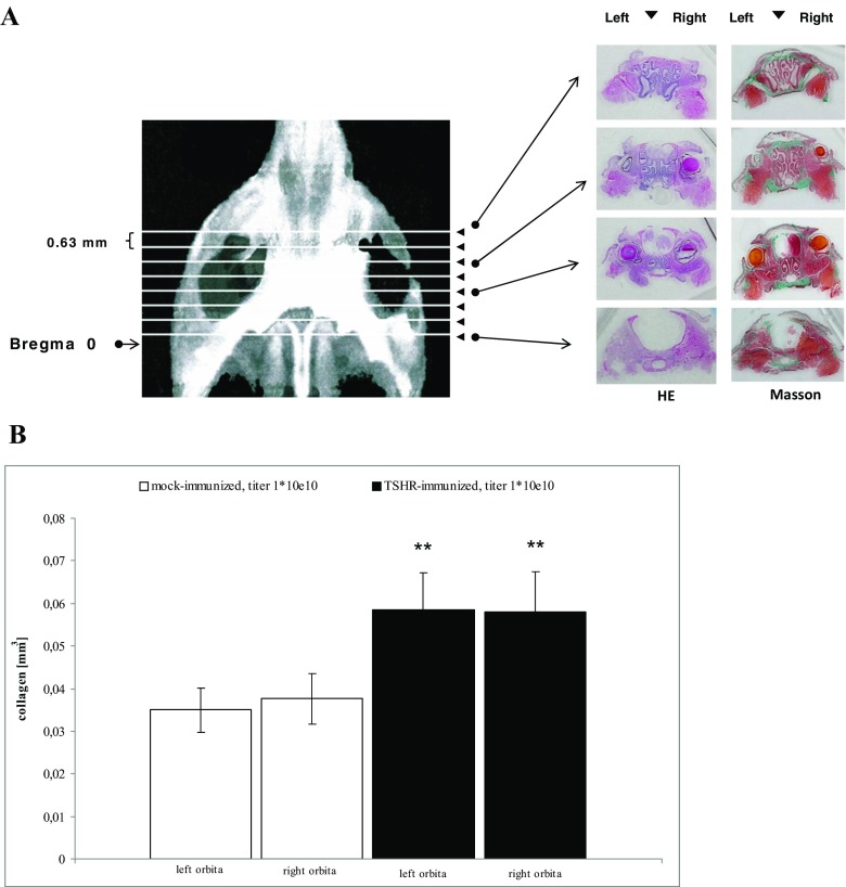
Fig. 1.
Histological investigation of orbital sections. a Representative macroscopic images. Representative images of coronary sections of a mouse orbita and neighbouring tissues. The sections were taken at defined distances from the mouse bregma. Interstitial connective tissue/fibrosis was then stained in green (Masson’s trichrome stain). For clarity, both HE-stained sections (left panels) and Masson’s stained sections (right panels) are shown next to each other. b Effect on digitised analysis of retroorbital tissue. The effects on severity of retro-orbital fibrosis were evaluated in histological sections. The measurements were carried out in immunised mice treated by either four weekly injections with control Ad-GFP (“mock-immunised”) or four weekly injections with Ad-TSHR (“Graves’ diesease”). N = 10 mice were investigated in each group. The mean total fibrosis volumes of each right and left orbita, as assessed by digitised image analysis of all sections, and consecutive integrations, are shown with SEM. Differences between groups were tested by ANOVA, *p < 0.05 compared to the TSHR-immunised group

